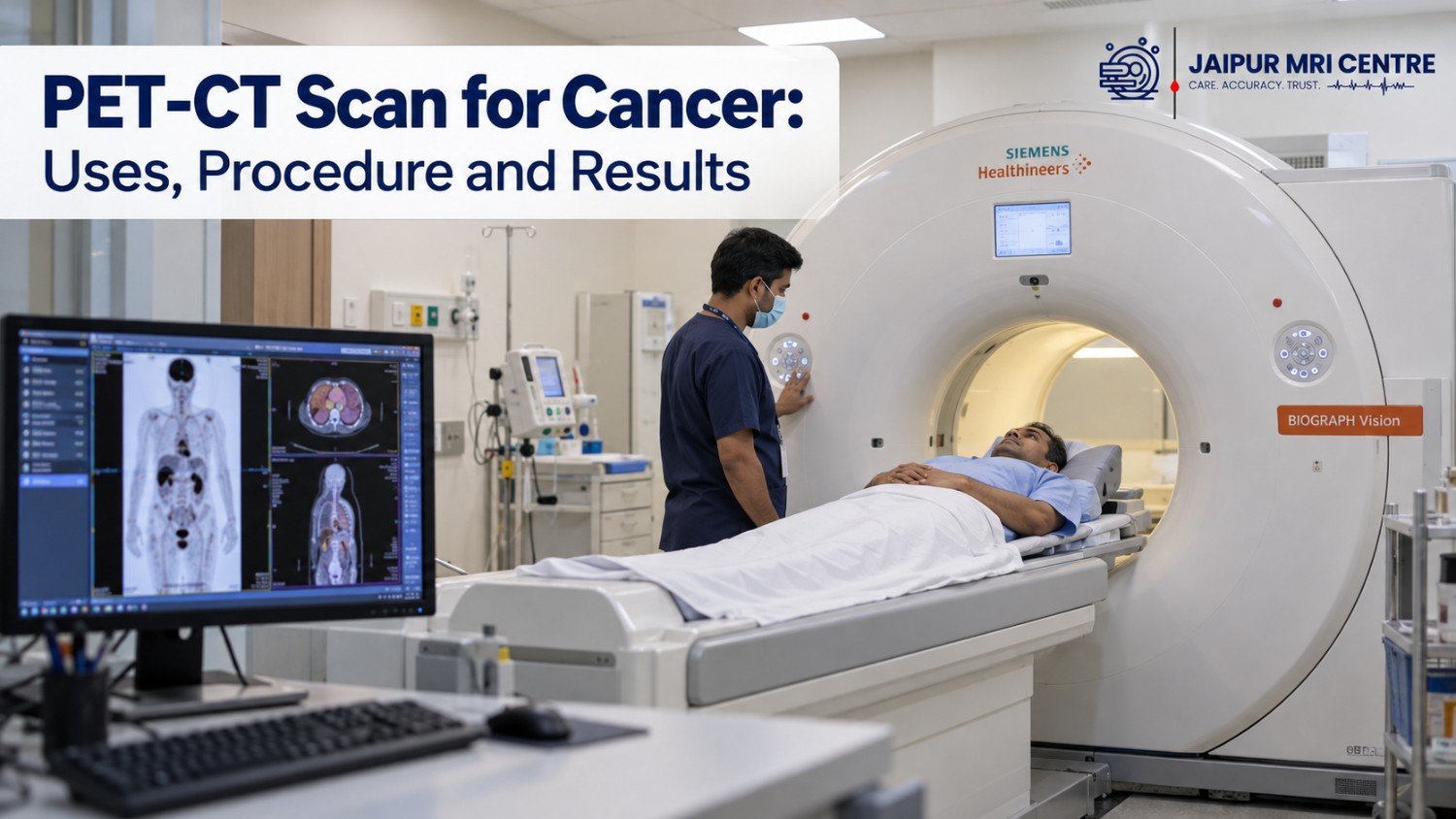
A cancer diagnosis is a life-changing event that raises a multitude of questions, concerns and decisions. In the modern era of oncology, precision is everything. Physically locating a tumor is not enough—doctors need to know how the cells in the tumor are functioning on a molecular and metabolic level.
This is where advanced nuclear medicine imaging can help. One of the most important and widely used diagnostic tools of the day is the PET-CT scan for cancer. A PET-CT scan offers a holistic view of cellular metabolism across the whole body by combining functional metabolic imaging with high-resolution anatomical mapping.
Preparing for a PET-CT scan can feel confusing for a patient and their family, but with knowledge of what a PET-CT scan is, why it may be ordered, how the procedure works and what the results mean, the medical process can be navigated more confidently and with greater clarity.
PET-CT is a combined imaging technique that combines two types of imaging into a single combined imaging session:
PET (Positron Emission Tomography): This is an imaging technique which assesses metabolic activity in the body. A tiny safe dose of radioactive tracer (usually Fluorodeoxyglucose, or FDG, a glucose analogue labelled with radioactive fluorine-18) is given before the scan.
Cancer cells grow quickly and take in a lot of energy to grow, which means they use more glucose than normal healthy cells. These are the areas where the PET scanner can see the uptake and are identified as “hot spots” of metabolism.
CT (Computed Tomography): This gives detailed anatomical mapping with the help of special X-ray machines. Produces images of blood vessels, organs and bones in cross-sectional 3D.
PET-CT has transformed cancer care throughout the cancer journey. There are a number of main reasons oncologists use this imaging test:
Once a patient has been diagnosed with cancer, the extent of the disease (the stage) will guide the overall treatment. Physicians are able to see primary tumors and small deposits of disease in distant lymph nodes, bones, liver, lungs or other organs across the entire body with a PET-CT scan, which conventional x-rays or ultrasounds may miss. Very small lesions can still be missed, so the scan is always read alongside the rest of your tests.
The standard imaging methods used (such as standard CT or MRI) do show the size of the tumor and whether it is shrinking or not. But sometimes after a successful treatment, the size of the tumour may not change and instead, it may be full of dead scar tissue or inactive cells. The PET-CT scan for cancer is not based on size but on metabolic activity. The absence of a “metabolic hot spot” suggests a good response to treatment, meaning the tumour cells are likely no longer active, although some structural mass may still be present. Your oncologist will interpret this together with your other findings.
A PET-CT scan is a highly useful tool to find a recurrence early if the patient has finished cancer treatment and blood tests or symptoms indicate the cancer has “come back”. It can often help distinguish scar tissue from active recurrent tumour growth.
PET-CT is used by the radiation oncologists to accurately delineate radiation therapy fields. They can determine the exact metabolic limits of a tumour, and focus high doses of radiation on active cancer cells without harming the surrounding healthy tissue.
FDG PET-CT is widely used for cancers such as lymphoma, lung, head and neck, oesophageal, colorectal, cervical cancer and melanoma. For some cancers, a different tracer works better: PSMA PET-CT is used for prostate cancer and DOTA-based scans for neuroendocrine tumours. Your doctor will decide whether PET-CT is right for your type and stage of cancer.
Booking a PET-CT scan in Jaipur?
Tell us your scan type and we will share partner-centre rates and the earliest available slots, so you can compare before you book. The scan and report are done by the independent diagnostic centre.
Having a special scan is a frightening experience, but it is good to know what to expect to lower anxiety. Below are typical steps in the process:
Dietary Restrictions: Fasting is usually required for 4-6 hours prior to an FDG-PET scan. You can take only plain water; however, sugar, carbohydrates, caffeine and heavy exercise will affect your body’s ability to absorb glucose. Avoid chewing gum and strenuous exercise before the scan.
Medications: Let your medical providers know of all the prescription medicines, supplements or treatment of diabetes that you are taking. The blood sugar level should be kept in a stable condition prior to the injection of the tracer. Most centres need it to be below roughly 180-200 mg/dL (the exact limit varies by centre), and the scan may be rescheduled if it is higher. Do not change your medicines or insulin on your own; follow the instructions given by the diagnostic centre.
The Injection: A little amount of the radiotracer (FDG) is given by injection into a vein, usually in your arm.
Uptake Wait: This is a period of 45 to 60 minutes during which you will spend a quiet period in a private room in the dark. You need to be as still as possible during this period, and not read, use your phone to scroll (or talk too much). During this resting period, the radiotracer moves through the blood and is taken up by any tissues that have a high metabolic rate. You may be asked to empty your bladder before the scan begins.
The PET-CT scanner is a large, donut-shaped machine, like a conventional CT scanner.
Scanning: You will be placed comfortably on a table inside a scanner ring which slowly passes you through the ring. You will be required to lie still and breathe normally (or hold your breath briefly for some chest scans).
Time: The scan itself takes 20-40 minutes, with the appointment taking about 2-3 hours in total, including preparation and uptake.
You can usually eat and go back to your routine. Drinking plenty of water helps your body flush out the tracer. The centre may advise you to limit close contact with infants and pregnant women for a few hours; follow the instructions you are given.
A PET-CT report is prepared by a nuclear medicine physician or radiologist after studying hundreds of images. It usually describes where abnormal metabolic activity (“hot spots”) was seen, how intense it is (often measured as an SUV value) and what the matching CT image shows at that spot.
Please do not rely on the report alone. Show it to your oncologist, who will explain what it means for your treatment plan.
The cost of PET-CT Scan is quite high, as it require state-of-the-art gamma-ray detectors and cyclotron facilities.
The total cost of a PET-CT scan for the whole body in Jaipur is likely to be in the range of ₹18,000, which depends on the tracer type being used (like standard FDG or specialized PSMA for prostate scans), the PET-CT facility, and the diagnostic centre.
The factors that impact the cost are:
The overall cost may be changed by type of tracer used: Standard glucose analogues (FDG) are common, while specialized tracers for a cancer diagnosis or infection might be different.
The rate of stand-alone advanced nuclear imaging centers is lower and more economical, as compared to those of multi–speciality corporate hospitals.
Extra protocols: If contrast material and/or targeted regional scans are performed as add-on to the whole-body protocol, the protocol may impact the overall cost.
For some patients, a complicated time can be made even more complicated by dealing with the hospital waiting lists, physician prescriptions, and certified centers when dealing with advanced imaging needs.
JAIPUR MRI CENTRE is a dedicated online appointment booking platform that helps patients to connect them with the best PET-CT scan centres in Jaipur. We do not perform scans or give medical advice; the scan and the report are done by connected partner diagnostic centres. Here is how we can help:
The PET-CT scan for cancer is one of the most important tools in cancer care today, providing a unique perspective on cell metabolism, cancer staging and the effectiveness of cancer treatment. It provides a molecular understanding of what occurs under the surface, enabling oncologists to create precise, effective therapeutic strategies.
If your doctor has suggested you have a PET-CT scan, remember early diagnosis and accurate staging is your best weapon in the battle against cancer.
Has your doctor advised a PET-CT scan?
Time matters in cancer staging and follow-up. Send us your doctor’s advice and your preferred date, and our team will help you find a partner centre and a suitable slot. Pre-scan instructions such as fasting and blood sugar are given by the diagnostic centre.
Jaipur MRI Centre is a booking platform. It does not perform scans or provide medical advice.
A PET CT scan will expose you to ionizing radiation from the radiotracer (e.g. FDG) and the CT. The dose is carefully controlled, however, to produce high-resolution images and be as low as is possible, but still diagnostic. The potential harm of one screening test is widely outweighed by the potential for benefit from accurate detection, staging or surveillance of cancer.
The most commonly used radiotracer in oncology is Fluorodeoxyglucose (FDG) , a modified version of sugar (glucose). Fasting for 4-6 hours prior to your appointment reduces your baseline blood sugar and insulin levels, which helps the cancer cells absorb the glucose.
This allows the radioactive tracer to be taken up by the active cells of the tumor, rather than competing with the sugars in a person’s bloodstream, and gives much clearer and more accurate images of the scan.
Yes, a diabetic patient can safely have a PET CT scan, however, it is essential that the patients’ blood sugar be controlled. If there is an elevation in blood sugar, the distribution of the tracer could be affected and the scan might not be as good.
If you take medicine for your diabetes, please let the staff at Jaipur MRI Centre know so we can give you special instructions about your medicine, insulin schedule and diet on the day of the MRI scan.
The PET-CT scan provides hundreds of high-resolution images of the body’s metabolism and structure that must be examined by an experienced nuclear medicine physician and radiologist.
These detailed reports and digital image plates are generally prepared and delivered in 24 – 48 hours. Jaipur MRI Centre also provides direct sharing of digital copies of your MRI scans on WhatsApp or email so that you can seek immediate advice from your oncologist.
Some of the more sophisticated imaging tests, such as PET-CT, may require a lot of cash. Jaipur MRI centre collaborates with registered nuclear medicine lab and premier diagnostics centres of Jaipur.
Our network provides us with exclusive discounted rates, ensuring that you can book without administrative hassles and at lower rates than the standard hospital charges.
Disclaimer: This content is for general information only and is not medical advice. Please consult your doctor before any test or treatment.


Need Help