Your doctor has told you to get a brain MRI, and your prescription reads either “MRI Brain – Plain” or “MRI Brain – Plain + Contrast.” Naturally, the first question is: what’s the difference, and which one do you actually need?
The reality is that both types of Brain MRIs are used for different medical reasons. Either one or the other isn’t necessarily better, but it will depend on the condition that your doctor is studying.
At JAIPUR MRI CENTRE, we assist patients in scheduling Brain MRI appointments at reputable diagnostic centres in Jaipur, which ensure state-of-the-art MRI machines, skilled radiologists and timely reporting of MRI reports. In this guide, we will discuss the difference between a Brain MRI with contrast and a Brain MRI without contrast (Plain MRI), when each is recommended, the benefits of each type, potential risks, and what you can expect during the procedure.

Get MRI Scan Price & Discount | Expert Help
| Questions | Non-contrast (Plain) MRI | MRI with Contrast |
|---|---|---|
| Injection involved? | No | Yes (gadolinium dye into a vein) |
| What’s good at | Brain anatomy, bleeds, acute stroke, structural problems | Highlighting abnormal tissue, blood supply, and disease activity |
| Common reasons | Headache, migraine, dizziness, seizures, acute stroke, head-injury follow-up, degenerative changes | Suspected tumor, infection/abscess, active MS lesions, post–brain-surgery review, suspected spread (metastasis), certain vascular problems |
| Typical extra time | — | Usually a few extra minutes for the injection and post-contrast images |
| Who decides | Your treating doctor/radiologist | Your treating doctor/radiologist |
If your prescription only says “Plain,” your doctor has judged that anatomical images are sufficient for now. If it says “Plain + Contrast,” they want the additional detail that the dye provides. You generally cannot self-select between them — the indication drives the choice.
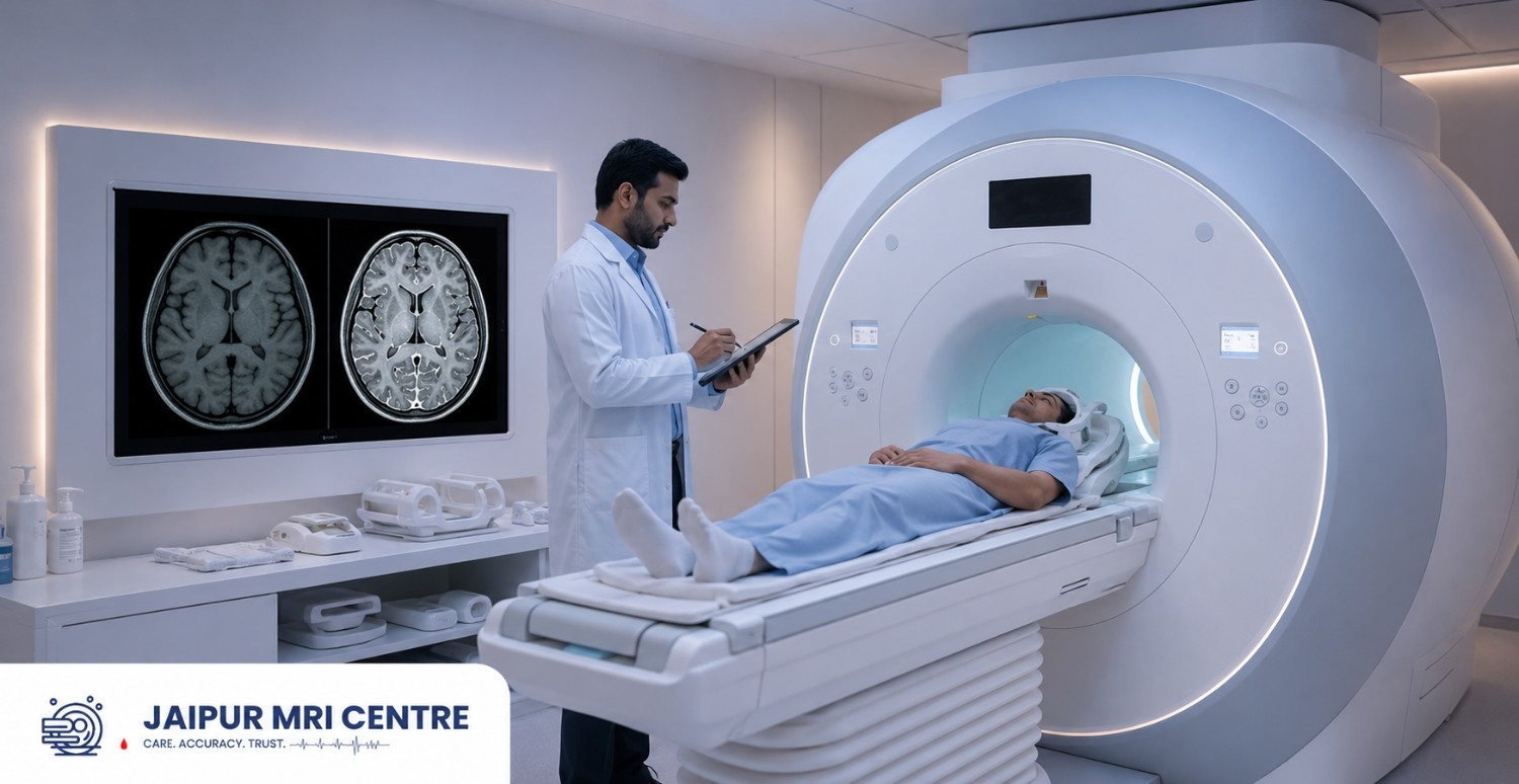
Brain MRI (Magnetic Resonance Imaging) is a safe diagnostic imaging procedure that produces high-resolution images of the brain and other neighbouring structures using a powerful magnetic field and radiofrequency waves.
An MRI scanner does not use X-rays or other types of radiation, making it a safer method of imaging, particularly for soft tissues, nerves and blood vessels, than a CT scan or X-ray.
A Brain MRI is often used to help diagnose:
Depending on the suspected condition, your doctor may recommend either a plain MRI or an MRI with contrast.

Avail up to 25% Off On MRI Scan
Brain MRI without contrast (Brain MRI – Plain MRI or Non-Contrast MRI) is an MRI that does not involve contrast dye.
The MRI is a machine that uses only magnetic fields to obtain very detailed images of the brain. In many neurological diseases, these images are enough to diagnose the disease. No contrast is needed making the procedure both simple and easy to prepare for, in most cases.
For most cases, the doctor typically orders a plain Brain MRI when:
When doctors need to get a detailed image of the anatomy of the brain, a non-contrast MRI may be the first imaging test ordered.
Brain MRI with contrast means that before or during the MRI, gadolinium-based contrast agent (a special dye) is placed into a vein.
The contrast gets carried throughout the body and helps make certain tissues, blood vessels and abnormal regions that you might not see well on a regular MRI.
It is not a replacement of MRI, but rather it adds to certain structures so that the radiologist can examine those in more detail. Contrast MRI is recommended when more information is needed than what is provided by a traditional MRI.
Doctors might want a contrast-enhanced brain MRI if they think they are encountering a condition that needs a detailed view of any abnormal tissue or blood supply.
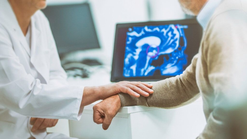
Image showing while the process of Brain MRI Scan Process by patient.
These include:
Contrast is used to make it easier to see if the tumour is different from the normal brain tissue. It also helps doctors to determine the size, location, blood supply, and extent of the tumour.
Also check, What to Expect During an MRI Scan: A Patient’s Complete Guide
After contrast, conditions like abscesses, complications caused by meningitis and some inflammatory disorders become more apparent.
This is why MS can appear on both types of scan. A plain MRI can detect MS lesions, but contrast shows whether a lesion is active (currently inflamed) or old and inactive important information for tracking the disease.
Patients who have had brain surgery might need to have contrast MRI to check for healing or recurrence of disease.
Contrast can help detect the presence of any secondary tumours which may have spread to the brain from the body.
For some aneurysms or inflammatory conditions of the vessels, contrast-enhanced imaging — or a related technique called MR angiography (MRA) , which images blood vessels — may be used for a fuller assessment.
The choice is not random, and it isn’t about cost or preference. Your doctor weighs:
If you’re unsure why a particular scan was chosen, it’s completely reasonable to ask your doctor to explain the reason in your specific case.
For most people, modern gadolinium-based contrast agents are considered very safe. Serious allergic reactions are uncommon, and the majority of patients experience no side effects.
Some people may notice:
These effects usually resolve quickly.
However, contrast may not be suitable for everyone.
Your doctor may avoid contrast if you:
Always inform your healthcare provider about your medical history before undergoing a contrast MRI.
Also check, Feeling Anxious? How to Manage MRI Claustrophobia & Panic During Scans

This step applies to both plain and contrast scans, and it matters because the MRI magnet is always on. Before your scan, tell the center if you have:
The center will give you a safety questionnaire. Answer it Honestly and completely some implants are MRI-safe and some are not, and the staff needs this information to keep you safe. You will also be asked to remove metal objects, jewelry, and certain clothing.
Once at the diagnostic centre, the necessary formalities will be carried out before you enter the MRI room.
For a plain MRI:
For a contrast MRI:
During the scan, you will have to lie on the MRI table in a comfortable position and the table will slide into the scanner.
Stay still-try not to move as this will cause the images to become blurred. The machine may make a loud knocking noise when scanning and earplugs or headphones are typically offered to make the process more comfortable.
Yes. If the MRI is normal, then you will be able to resume normal activities immediately. To help the contrast dye be excreted naturally by the kidneys, patients are advised to drink lots of water throughout the day after the MRI.
There are no restrictions following the scan unless your doctor recommends otherwise. Patients can schedule their Brain MRI at JAIPUR MRI CENTRE at various centres in Jaipur with top radiologists and advanced MRI machines at transparent pricing.
For neurological imaging, image quality and accurate reporting will be of prime importance.
Check below these why patient choose us
We want the MRI experience to be easy, dependable and convenient from appointment to report pick-up.
Brain MRI with contrast and Brain MRI w/o contrast both provide valuable diagnostic information; however, for different uses. A simple MRI can be used to help diagnose many neurological disorders, seizures, dizziness and headaches. A contrast-enhanced MRI is advised if a more detailed assessment is required, such as for tumours, infections, inflammation or some vascular disorders.
This should always be based upon your doctor’s recommendation and your medical condition. The right scan performed at a reputable diagnostic facility can help to make a proper diagnosis and aid in proper treatment.
Brain MRI in Jaipur is available at the JAIPUR MRI CENTRE, where you can easily find appointments at the diagnostic centres that employ the latest imaging technology, have experienced radiologists, and ensure reliable reporting.
Neither is universally better. They serve different purposes. A plain MRI answers many structural and neurological questions; a contrast MRI is added when your doctor needs to study abnormal tissue, blood supply, or disease activity more closely.
You may feel a brief pinch when the IV is placed and a cool sensation as the dye goes in. Significant pain is not expected.
For most people, yes. Reactions are uncommon and usually mild. Gadolinium can be retained in the body in small amounts, but this has not been linked to harm in people with normal kidney function. Tell your team about any kidney disease, pregnancy, or past contrast reaction.
In most cases, yes — there are usually no restrictions after a routine brain MRI. Follow any specific advice your doctor or the center gives you.
Often, yes, with the right agent and precautions. Modern contrast agents are considered low-risk even in reduced kidney function, but your radiologist will assess this individually, so always disclose any kidney disease.
Yes. Only a very small amount of contrast enters breast milk, and breastfeeding can usually continue normally.
This article is for educational purposes only and should not replace professional medical advice, diagnosis, or treatment. Always consult your doctor or radiologist for medical concerns.
From brain and spine to joint and full body imaging, access safe and precise MRI diagnostics with quick appointment availability.
Need Help